|
8:00 am
|
Welcome and Opening Remarks - Ontario Association of Medical Radiation Sciences
|
|
|
8:10
am
|
Update on Breast Cancer Screening in Canada
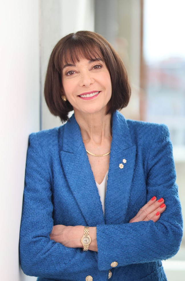
Dr. Paula Gordon, OC, OBC. MD, FRCPC, FSBI, Clinical Professor, University of British Columbia, Founding Medical Director, Sadie Diamond Breast Program, BC Women's Hospital
Dr. Gordon is a Clinical Professor of Radiology at the University of British Columbia. Her research was the first to show that ultrasound could find small, non-palpable cancers not seen on mammograms in women with dense breasts. This has led to a paradigm shift in screening women with dense breasts that started in the USA in 2009, and is now spreading globally. She volunteers as Medical Advisor to DenseBreastsCanada.ca and DenseBreast-Info.org, and as Vice President of the Board of the Canucks for Kids Fund. Among other recognitions of her contributions to the field of breast imaging, she was invested in the Order of British Columbia in 2013, and appointed as an Officer of the Order of Canada in 2022.
At the conclusion of Dr. Gordon's presentation, attendees will understand the benefits of early detection of breast cancer and the risks of screening. They will also understand the risks associated with dense breasts and the importance of women being notified of their own breast density and will be familiar with what breast screening is currently available in Ontario, and what changes the province has committed to making.
|
|
|
9:00 am
|
Updates to the Ontario Breast Screening Program
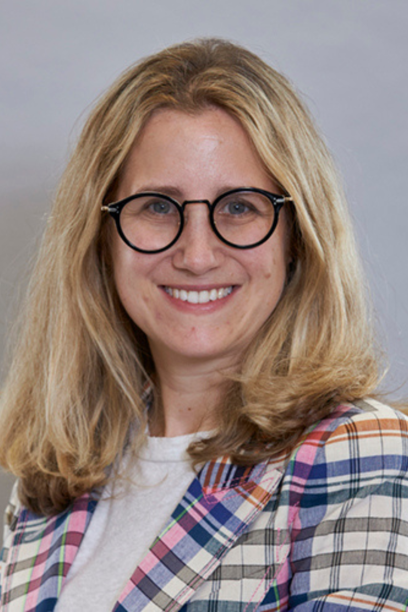
Dr. Samantha Fienberg, MD, FRCPC, MBA, Clinical Lead for the Ontario Breast Screening Program and Breast Imaging Lead and Staff Radiologist at Lakeridge Health
Dr. Samantha Fienberg is the Clinical Lead for the Ontario Breast
Screening Program. She is also the Breast Imaging Lead and staff
radiologist at Lakeridge Health. Dr. Fienberg has a clinical interest in
improving patient access, and improving quality of care within medical imaging. She also has a clinical interest in incorporating advanced technologies into clinical practice in the community setting. Dr. Fienberg is passionate about providing high quality breast cancer imaging in the community. Given current healthcare challenges, she appreciates the opportunity to discuss these issues.
In this presentation, Dr. Fienberg will provide an overview of the function of the OBSP and its basic structure as well as an update on the upcoming expansion of the Ontario Breast Screening Program to include people ages 40 to 49. She will also discuss supplemental breast screening.
|
|
|
9:30
am
|
Unveiling the Realms of Mammographic Guided Procedures
Dr. Priscila Crivellaro, MD, Clinician, Assistant Professor, Western University, St. Joseph's Health Care
Dr. Priscila Crivellaro is an assistant professor in the Department of
Medical Imaging at Western University in London, Ontario, Canada.
She completed three years of women’s imaging fellowship training at the University of Toronto, and has more than nine years of experience in breast imaging. Her research interests are improvements in patient outcomes in the breast imaging field, artificial intelligence in radiology, global health, and medical continued education.
Mammographic-guided procedures encompass a critical area of breast imaging, with variable techniques implemented, including stereotactic technique, tomosynthesis and contrast-enhanced mammography-guided procedures.
By the end of this lecture, you will be able to:
- Identify the clinical indications for each specific modality of
mammographic-guided procedures.
- Analyze the steps in performing the distinct mammographic-guided procedures.
- Discuss these procedures' challenges and limitations to understand the outcomes better.
Presentation #2 - Elastography on Breast Imaging: Why, When and How
The main objectives for this presentation include:
- Providing a review the basics of this technique and how to perform the examination.
- Identifying the scenarios where elastography can affect the management and improve diagnostic accuracy.
- Internalize the main findings for imaging interpretation and common pitfalls.
|
|
|
10:20
am
|
Break
|
|
|
10:30
am
|
Current State of the Art Magnetic Resonance Imaging of Breast Cancer
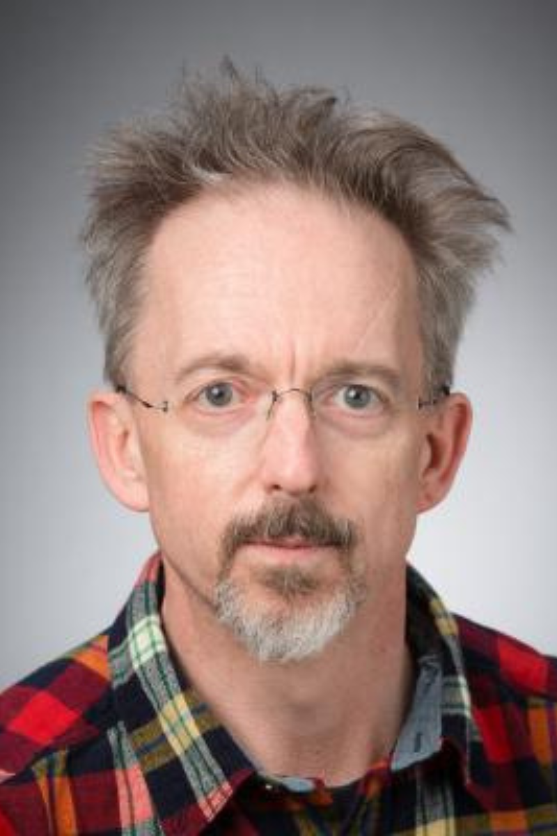
Michael Noseworthy, PhD, PEng, MSc, Co-Director, McMaster School of Biomedical Engineering, Special professional staff in Radiology and Nuclear Medicine, St. Joseph's Health Care
Dr. Michael D. Noseworthy, received a M.Sc. for work in the evaluation of anaesthetic hepatotoxicity using MRI, transmission electron microscopy (TEM) and in vivo 31P-NMR. He then obtained a PhD (1997) specializing in applications of MRI/MRS and electron paramagnetic resonance (EPR) methods to assess free radical induced brain damage. From 1997-1999 was a postdoctoral fellow in Imaging Physics, Sunnybrook Health Sciences Centre (Toronto) working on the evaluation of tissue microvasculature through development of correlative MRI and energy dispersive X-ray microanalysis (EDXS). From January 2000 to August 2003 he worked as a MRI physicist at The Hospital for Sick Children and University Health Network (UHN), Toronto, and was Assistant Professor in Medical Biophysics and Medical Imaging, University of Toronto. He was recruited to St. Joseph’s Healthcare and Brain - Body Institute, McMaster University in August 2003. Following 3 years as an Assistant Professor in Radiology and Medical Physics at McMaster, Dr. Noseworthy obtained a tenure-track assistant professor position in Electrical & Computer Engineering at McMaster University, where he currently resides as a full professor. Also at McMaster University he was the Co-Director of the School of Biomedical Engineering (2010-2020) and still is Director of Imaging Physics and Engineering at the Imaging Research Centre, St. Joseph’s Healthcare, Hamilton. Dr. Noseworthy also has Special Professional Staff status at St. Joseph’s Healthcare in both Radiology and Nuclear Medicine. He is or has been funded by NSERC, CIHR, CFI and ORF, has published over 150 peer reviewed papers, 270 conference papers and had 75 graduate students and postdocs. Dr. Noseworthy is a member of the Professional Engineers of Ontario (PEO), International Society for Magnetic Resonance in Medicine (ISMRM) and European Society for Magnetic Resonance in Medicine and Biology (ESMRMB).
In his presentation, Dr. Noseworthy will review what’s new in breast MRI acquisition and also image classification. These new approaches focus on machine learning, and more specifically deep learning. He will outline how these are improving dynamic contrast enhanced MRI (dceMRI) and diffusion weighted MRI of breast cancer.
By the end of this lecture, you will be able to:
- Identify what's new in breast MR imaging.
- Understand deep learning and how it is speeding up MRI acquisition.
- Identify how artificial intelligence is being used to assist in breast tumor classification.
|
|
|
11:00
am
|
Session
to be announced
Sarah
Alexander, Account Executive, Breast and Skeletal Health
Hologic
|
|
|
11:20
am
|
.png)
Ultrasound
Correlation of Mammographic Abnormalities
Dr. Ameya Madhav Kulkarni, Hamilton Health Sciences,
McMaster University
Dr.
Ameya Madhav Kulkarni is a staff Radiologist at Juravinski Hospital and
Cancer Centre in Hamilton. He completed his my fellowship in Breast and
Cross-Sectional Imaging from McMaster University in 2020. Dr. Kulkarni worked
as a staff radiologist in a teaching hospital in India and always wanted to
do more work in cancer imaging. He enjoys supervising and training fellows
and residents in hospital and is currently a fellowship co-ordinator at
Juravinski Hospital. His clinical interests are breast & body oncology
imaging and he continues to pursue research in Radiomics.
In
his talk, Dr. Kulkarni will cover the appearance of mammographic
abnormalities in ultrasound. He will also discuss indications of ultrasound
and the role of ultrasound in staging.
|
|
|
11:50
am
|
Lunch
|
|
|
12:20
pm
|
Automated Breast Ultrasound
Anju Tomar, MHA, RDMS, RVT, National Applications Director, Automated Breast Ultrasound (ABUS), GE HealthCare
In her current role, Anju lives and and breathes all things ABUS. She is extremely passionate about educating women about breast density and believes in early detection of cancer and making a difference in the lives of women with dense breast tissue. During her 22 years at GE, 10 of them have been spent gaining experience across everything it takes to build a successful breast imaging screening program, inclusive of: sales, clinical applications, marketing, product development, program implementation, education, IT/Workflow, service, and project management. Her passion and purpose is to make ultrasound and adjunct screening modality accessible to every woman that needs it.
In this presentation, Anju will discuss personalized screening for women with dense breasts.
|
|
|
12:40
pm
|

Breast Imaging: The Patient Experience
Lyndsay Simmons, MRT (MR) (R), PhD Candidate|Radiation Sciences, Centre for Integrated and Advanced Medical Imaging, Mohawk College
Lyndsay Simmons graduated from Charles Sturt University and holds a Masters degree in Medical Radiation Sciences with a speciality in Computed Tomography. She is also a certified MRI technologist and is currently working towards her PhD in Radiation Sciences at McMaster University. Lyndsay has worked as a college professor in the school of Medical Imaging at Mohawk College since 2008 and supports both academic and research activity at the Centre for Integrated and Advanced Medical Imaging.
In this presentation, attendees will learn about a patient's imaging experience and high-risk patients, the integral role of patient communication and booking, as well as sensitivity and specifically breast imaging.
|
|
|
1:30
pm
|
Session
to be announced
Melissa Reeve, Women's Health Product Specialist
Siemens Canada
|
|
|
2:00
pm
|
Interactive Hands-on Demonstrations
Participate in hands-on demonstrations of the latest mammography and ultrasound equipment from Siemens Healthineers, Hologic and GE HealthCare. Step in to the Siemens Healthineers demo truck for the latest in breast imaging technology.
|
|
|
5:00
pm
|
Closing
Remarks
Ontario Association of Medical Radiation Sciences
|
|